Monday, October 24, 2011

MMP3 and lung fibrosis

An increase in MMP3 was found in IPF lung tissue. Mmmp3–/– mice were protected from bleomycin induced lung fibrosis, through beta-catenin mediated EMT.


References:
Am J Pathol. 2011 Oct;179(4):1733-45. Epub 2011 Aug 24.
Matrix metalloproteinase 3 is a mediator of pulmonary fibrosis.
Yamashita CM, Dolgonos L, Zemans RL, Young SK, Robertson J, Briones N, Suzuki T, Campbell MN, Gauldie J, Radisky DC, Riches DW, Yu G, Kaminski N, McCulloch CA, Downey GP.
Source
Department of Medicine, University of Western Ontario, London, Ontario, Canada; Division of Pulmonary and Critical Care Medicine, Departments of Medicine and Pediatrics, National Jewish Health, Denver, Colorado.
Abstract
Idiopathic pulmonary fibrosis (IPF) may be triggered by epithelial injury that results in aberrant production of growth factors, cytokines, and proteinases, leading to proliferation of myofibroblasts, excess deposition of collagen, and destruction of the lung architecture. The precise mechanisms and key signaling mediators responsible for this aberrant repair process remain unclear. We assessed the importance of matrix metalloproteinase-3 (MMP-3) in the pathogenesis of IPF through i) determination of MMP-3 expression in patients with IPF, ii) in vivo experiments examining the relevance of MMP-3 in experimental models of fibrosis, and iii) in vitro experiments to elucidate possible mechanisms of action. Gene expression analysis, quantitative RT-PCR, and Western blot analysis of explanted human lungs revealed enhanced expression of MMP-3 in IPF, compared with control. Transient adenoviral vector-mediated expression of recombinant MMP-3 in rat lung resulted in accumulation of myofibroblasts and pulmonary fibrosis. Conversely, MMP-3-null mice were protected against bleomycin-induced pulmonary fibrosis. In vitro treatment of cultured lung epithelial cells with purified MMP-3 resulted in activation of the β-catenin signaling pathway, via cleavage of E-cadherin, and induction of epithelial-mesenchymal transition. These processes were inhibited in bleomycin-treated MMP-3-null mice, as assessed by cytosolic translocation of β-catenin and cyclin D1 expression. These observations support a novel role for MMP-3 in the pathogenesis of IPF, through activation of β-catenin signaling and induction of epithelial-mesenchymal transition.
Copyright © 2011 American Society for Investigative Pathology. Published by Elsevier Inc. All rights reserved.
PMID: 21871427 [PubMed - in process] PMCID: PMC3181358 [Available on 2012/10/1]

Thursday, October 20, 2011

Plasma proteins predict IPF

With a cohort of 241 IPF patients, a recent study found that high concentrations of MMP7, ICAM1, IL8, VCAM1, and S100A12 predicted poor overall survival, poor transplant free survival and poor progression free survival in the derivation cohort.


References:
Am J Respir Crit Care Med. 2011 Oct 20. [Epub ahead of print]
Peripheral Blood Proteins Predict Mortality in Idiopathic Pulmonary Fibrosis.
Richards TJ, Kaminski N, Baribaud F, Flavin S, Brodmerkel C, Horowitz D, Li K, Choi J, Vuga LJ, Lindell KO, Klesen M, Zhang Y, Gibson KF.
Source
Medicine/PACCM, University of Pittsburgh, Pittsburgh, Pennsylvania, United States.
Abstract
Introduction/

BACKGROUND:
Idiopathic pulmonary fibrosis (IPF) is a lethal lung disease of unknown etiology with a variable and unpredictable course. The aim of this study was to identify and validate plasma proteins that are predictive of outcome in IPF.

METHODS:
Plasma samples were available for 241 IPF patients (140 - derivation and 101 validation). In derivation cohort, concentrations of 92 proteins were analyzed using a multiplex bead-based immunoassay and concentrations of MMP7, MMP1, and SPD were assessed by ELISA. In the validation cohort concentrations of ICAM1, IL8, VCAM1 were assessed by bead-based multiplex assay, and S100A12 and MMP7 by ELISA. Associations of biomarkers with mortality, transplant free survival, and disease progression were tested in derivation and validation cohorts using nonparametric methods of survival analysis and the Cox proportional hazards model, and an integrated risk prediction score was derived and tested.

RESULTS:
High concentrations of MMP7, ICAM1, IL8, VCAM1, and S100A12 predicted poor overall survival, poor transplant free survival and poor progression free survival in the derivation cohort. In the independent validation cohort high concentrations of all five were predictive of poor transplant free survival, MMP7, ICAM1, and IL8 of overall survival and ICAM1 of poor progression free survival. The personal clinical and molecular mortality prediction index (PCMI) derived in the derivation cohort was highly predictive of mortality in the validation cohort.

CONCLUSIONS:
Our results suggest that plasma proteins should be evaluated as a tool for prognosis determination in prioritization of patients for lung transplantation and stratification in drug studies.

PMID: 22016448 [PubMed - as supplied by publisher]

Wednesday, October 19, 2011

Annexin A1 and lung fibrosis

Annexin A1 is upregulated in epithelial cells and inflammatory cells. In AnxA1 null mice, there were increased inflammation and fibrosis after bleomycin. There was also an increase in TGFbeta, IFN-gamma, and TNF-a.
The study suggests a protective role for endogenous Annexin A1 in lung fibrosis.

References:
BMC Immunol. 2011 Oct 19;12(1):59. [Epub ahead of print]
Endogenous annexin A1 counter-regulates bleomycin-induced lung fibrosis.
Damazo AS, Sampaio AL, Nakata CM, Flower RJ, Perretti M, Oliani SM.
Abstract
ABSTRACT:

BACKGROUND:
The balancing functions of pro/anti-inflammatory mediators of the complex innate responses have been investigated in a variety of experimental inflammatory settings. Annexin-A1 (AnxA1) is one mediator of endogenous anti-inflammation, affording regulation of leukocyte trafficking and activation in many contexts, yet its role in lung pathologies has been scarcely investigated, despite being highly expressed in lung cells. Here we have applied the bleomycin lung fibrosis model to AnxA1 null mice over a 21-day time-course, to monitor potential impact of this mediator on the control of the inflammatory and fibrotic phases.

RESULTS:
Analyses in wild-type mice revealed strict spatial and temporal regulation of the Anxa1 gene, e.g. up-regulation in epithelial cells and infiltrated granulocytes at day 7, followed by augmented protein levels in alveolar macrophages by day 21. Absence of AnxA1 caused increases in: i) the degree of inflammation at day 7; and ii) indexes of fibrosis (assessed by deposition of hydroxyproline in the lung) at day 7 and 21. These alterations in AnxA1 null mice were paralleled by augmented TGF-beta1, IFN-gamma and TNF-alpha generation compared to wild-type mice. Finally, treatment of wild type animals with an AnxA1 peptido-mimetic, given prophylactically (from day 0 to 21) or therapeutically (from day 14 onward), ameliorated both signs of inflammation and fibrosis.

CONCLUSION:
Collectively these data reveal a pathophysiological relevance for endogenous AnxA1 in lung inflammation and, more importantly, fibrosis, and may open new insights for the pharmacological treatment of lung fibrosis.

PMID: 22011168 [PubMed - as supplied by publisher]

Friday, October 14, 2011

Tight junction protein Claudin-5 and lung fibrosis


Am J Physiol Lung Cell Mol Physiol. 2011 Oct 14. [Epub ahead of print]
Altered expression of tight junction molecules in alveolar septa in lung injury and fibrosis.
Ohta H, Chiba S, Ebina M, Furuse M, Nukiwa T.
Source
1Tohku University Graduate school of Medicine.
Abstract
The dysfunction of alveolar barriers is a critical factor in the development of lung injury and subsequent fibrosis, but the underlying molecular mechanisms remain poorly understood. To clarify the pathogenic roles of tight junctions in lung injury and fibrosis, we examined the altered expression of claudins, the major components of tight junctions, in the lungs of disease models with pulmonary fibrosis. Among the 24 known claudins, claudin-1, claudin-3, claudin-4, claudin-7, and claudin-10 were identified as components of airway tight junctions. Claudin-5 and claudin-18 were identified as components of alveolar tight junctions and were expressed in endothelial and alveolar epithelial cells, respectively. In experimental bleomycin-induced lung injury, the levels of mRNA encoding tight junction proteins were reduced, particularly that of claudin-18. The integrity of the epithelial tight junctions was disturbed in the fibrotic lesions 14 days after the intraperitoneal instillation of bleomycin. These results suggest that bleomycin mainly injured alveolar epithelial cells and impaired alveolar barrier function. In addition, we analyzed the influence of transforming growth factor-β (TGF-β), a critical mediator of pulmonary fibrosis that is upregulated after bleomycin-induced lung injury, on tight junctions in vitro. The addition of TGF-β decreased the expression of claudin-5 in human umbilical vein endothelial cells (HUVECs) and disrupted the tight junctions of epithelial cells (A549). These results suggest that bleomycin-induced lung injury causes pathogenic alterations in tight junctions and that such alterations seem to be induced by TGF-β.

PMID: 22003091 [PubMed - as supplied by publisher]

Tuesday, October 11, 2011

SPARC in lung fibrosis

A recent study with bone marrow chimera mice suggested that, bleomycin induces inflammation and fibrosis in wild type through TNF synthesis that triggers TGF-β release; TGF-β promotes fibroblasts deposition and regulates TNF synthesis from macrophages.

B: In WT>SPARC KO chimeras, despite a normal parenchyma inflammation, collagen deposition by fibroblasts is greatly reduced and results in milder fibrosis.

C: In SPARC KO>WT chimeras, the inability of SPARC KO macrophages to down-modulate TNF production in response to TGF-β results in exaggerated and persistent inflammation and severe fibrosis.


Reference
Am J Pathol. 2011 Oct 11. [Epub ahead of print]
SPARC Oppositely Regulates Inflammation and Fibrosis in Bleomycin-Induced Lung Damage.
Sangaletti S, Tripodo C, Cappetti B, Casalini P, Chiodoni C, Piconese S, Santangelo A, Parenza M, Arioli I, Miotti S, Colombo MP.
Source
Molecular Immunology Unit, Department of Experimental Oncology and Molecular Medicine, Fondazione IRCCS Istituto Nazionale Tumori, Milan, Italy.
Abstract
Fibrosis results from inflammatory tissue damage and impaired regeneration. In the context of bleomycin-induced pulmonary fibrosis, we demonstrated that the matricellular protein termed secreted protein acidic and rich in cysteine (SPARC) distinctly regulates inflammation and collagen deposition, depending on its cellular origin. Reciprocal Sparc(-/-) and wild-type (WT) bone marrow chimeras revealed that SPARC expression in host fibroblasts is required and sufficient to induce collagen fibrosis in a proper inflammatory environment. Accordingly, Sparc(-/-) WT chimeras showed exacerbated inflammation and fibrosis due to the inability of Sparc(-/-) macrophages to down-regulate tumor necrosis factor production because of impaired responses to tumor growth factor-β. Hence, the use of bone marrow cells expressing a dominant-negative form of tumor growth factor-β receptor type II under the monocyte-specific CD68 promoter, as a decoy, phenocopied Sparc(-/-) donor chimeras. Our results point to an unexpected dual role of SPARC in oppositely influencing the outcome of fibrosis.
Copyright © 2011 American Society for Investigative Pathology. Published by Elsevier Inc. All rights reserved.
PMID: 22001347 [PubMed - as supplied by publisher]

Wednesday, October 5, 2011

CD69 and lung fibrosis

CD69-/- mice exhibited reduced inflammation and fibrosis.

Reference:
Respir Res. 2011 Oct 5;12(1):131. [Epub ahead of print]
Attenuation of lung inflammation and fibrosis in CD69-deficient mice after intratracheal bleomycin.
Yamauchi K, Kasuya Y, Kuroda F, Tanaka K, Tsuyusaki J, Ishizaki S, Matsunaga H, Iwamura C, Nakayama T, Tatsumi K.
Abstract
ABSTRACT:

BACKGROUND:
Cluster of differentiation 69 (CD69), an early activation marker antigen on T and B cells, is also expressed on activated macrophages and neutrophils, suggesting that CD69 may play a role in inflammatory diseases. To determine the effect of CD69 deficiency on bleomycin(BLM)-induced lung injury, we evaluated the inflammatory response following intratracheal BLM administration and the subsequent fibrotic changes in wild type (WT) and CD69-deficient (CD69-/-) mice.

METHODS:
The mice received a single dose of 3 mg/kg body weight of BLM and were sacrificed at 7 or 14 days post-instillation (dpi). Lung inflammation in the acute phase (7 dpi) was investigated by differential cell counts and cytokine array analyses of bronchoalveolar lavage fluid. In addition, lung fibrotic changes were evaluated at 14 dpi by histopathology and collagen assays. We also used reverse transcription polymerase chain reaction to measure the mRNA expression level of transforming growth factor beta1 (TGF-beta1) in the lungs of BLM-treated mice.

RESULTS:
CD69-/- mice exhibited less lung damage than WT mice, as shown by reductions in the following indices: (1) loss of body weight, (2) wet/dry ratio of lung, (3) cytokine levels in BALF, (4) histological evidence of lung injury, (5) lung collagen deposition, and (6) TGF-beta1 mRNA expression in the lung.

CONCLUSION:
The present study clearly demonstrates that CD69 plays an important role in the progression of lung injury induced by BLM.

PMID: 21970554 [PubMed - as supplied by publisher] Free

Sunday, October 2, 2011

Immunoglobulin free light chains are increased in hypersensitivity pneumonitis and IPF

from PLoSOne
PLoS One. 2011;6(9):e25392. Epub 2011 Sep 28.
Immunoglobulin free light chains are increased in hypersensitivity pneumonitis and idiopathic pulmonary fibrosis.
Groot Kormelink T, Pardo A, Knipping K, Buendía-Roldán I, García-de-Alba C, Blokhuis BR, Selman M, Redegeld FA.
Source
Division of Pharmacology and Pathophysiology, Utrecht Institute for Pharmaceutical Sciences, Faculty of Science, Utrecht University, Utrecht, The Netherlands.
Abstract
BACKGROUND:
Idiopathic pulmonary fibrosis (IPF), a devastating lung disorder of unknown aetiology, and chronic hypersensitivity pneumonitis (HP), a disease provoked by an immunopathologic reaction to inhaled antigens, are two common interstitial lung diseases with uncertain pathogenic mechanisms. Previously, we have shown in other upper and lower airway diseases that immunoglobulin free light chains (FLCs) are increased and may be involved in initiating a local inflammation. In this study we explored if such a mechanism may also apply to HP and IPF.

METHODS:
In this study we examined the presence of FLC in serum and BAL fluid from 21 IPF and 22 HP patients and controls. IgG, IgE and tryptase concentrations were measured in BAL fluid only. The presence of FLCs, plasma cells, B cells and mast cells in lung tissue of 3 HP and 3 IPF patients and 1 control was analyzed using immunohistochemistry.

RESULTS:
FLC concentrations in serum and BAL fluid were increased in IPF and HP patients as compared to control subjects. IgG concentrations were only increased in HP patients, whereas IgE concentrations were comparable to controls in both patient groups. FLC-positive cells, B cells, plasma cells, and large numbers of activated mast cells were all detected in the lungs of HP and IPF patients, not in control lung.

CONCLUSION:
These results show that FLC concentrations are increased in serum and BAL fluid of IPF and HP patients and that FLCs are present within affected lung tissue. This suggests that FLCs may be involved in mediating pathology in both diseases.

PMID: 21980441 [PubMed - in process] PMCID: PMC3182208

Sunday, September 25, 2011

BIBF 1120 for IPF

BIBF 1120 is developed  by Boehringer Ingelheim. It is a tyrosine kinase inhibitor targeting multiple tyrosine kinase receptors including platelet-derived growth factor receptors (PDGFR) α and β, vascular endothelial growth factor receptors
(VEGFR) 1, 2, and 3, and fibroblast growth factor receptors (FGFR) 1, 2, and 3.

A recent 12-month, randomized, double-blind, placebo-controlled, phase 2 trial (To Improve Pulmonary Fibrosis with BIBF 1120 [TOMORROW]) with 4 doses (50 mg once a day, 50 mg twice a day, 100 mg twice a day, or 150 mg twice a day) may have some positive effect (N = 428 patients). Although highest dose was associated with significant side effects, 150 mg twice a day was associated with a trend toward a reduction in the decline in lung function, with fewer acute exacerbations and preserved quality of life. Although the differences in primary end point among the study groups were not statistically significant, the benefits observed in the study were indeed clinically relevant.

It is encouraging that the trial was, at least, not another failed IPF trial, compared to others (pirfenidone, systemic glucocorticoids, azathioprine and cyclophosphamide, interferon-γ-1b, phosphodiesterase-5 inhibitors, or endothelin receptor antagonists).

I do hope that phase III trial gives more positive news. Since lower doses did not show much, I would guess that next  trial uses highest dose, 150 mg b.i.d.

References:
Richeldi L, et al. Efficacy of a tyrosine kinase inhibitor in idiopathic pulmonary fibrosis. N Engl J Med. 2011 Sep 22;365(12):1079-87. PubMed PMID: 21992121.
NEJM link: http://www.nejm.org/doi/full/10.1056/NEJMoa1103690.


Downey GP. Resolving the scar of pulmonary fibrosis. N Engl J Med. 2011 Sep
22;365(12):1140-1. PubMed PMID: 21992127.
NEJM link: http://www.nejm.org/doi/full/10.1056/NEJMe1108558.


Monday, September 12, 2011

FoxO3a and Lung Fibrosis

Dr. Henke and associates recent suggested that FoxO3a is largely inactive in IPF fibroblasts to promote IPF fibroblast proliferation. They previously demonstrated that lower Pten activity in IPF fibroblasts, higher PI3K-AKT activity on collagen matrix. In this study, the authors showed that phosphorylated, inactive, FoxO3 is higher, whereas total FoxO3 is lower, in IPF fibroblasts on collagen. They went on to show that p27 was lower in IPF.  They proposed model: 


Reference:
Nho RS, Hergert P, Kahm J, Jessurun J, Henke C. Pathological Alteration of
FoxO3a Activity Promotes Idiopathic Pulmonary Fibrosis Fibroblast Proliferation
on Type I Collagen Matrix. Am J Pathol. 2011 Sep 3. [Epub ahead of print] PubMed PMID: 21893017.

Friday, September 9, 2011

Human Lung Stem Cells?

Recently, Dr. Piero Anversa of Brigham and Women’s reported identification of human lung stem cells, appeared in May 12, 2011, issue of New England Journal of Medicine. The flow cytometric identification was based on c-kit antigen. The authors showed that the stem cells isolated were clonal and pluripotent. c-kit+ cells expressed KLF4, nanog,, oct4, and sox2. In a cryo injury mouse model, the authors showed that c-kit+  cells were, amazingly, able to promote lung regeneration!

In an editorial, Dr. Chapman of UCSF stated that "Kajstura et al. resets the starting point for clinical investigation into the role of stem cells and progenitor cells in lung disease". 

However, several scientists in the field were more skeptical. In Nature Medicine Community Corner, Dr. Stripp and Dr. Hogan of Duke questioned the experimental methodology (eg, RT-PCR and antibody staining) and proper controls.  They urged that more vigorous validation is needed. More importantly, the c-kit-CreER mice is needed to trace if the c-kit+ cells in the LUNG are pluripotent, hierarchical differentiation. Dr. Thannickal of UAB also suggested “The phenotype and fate of endogenous lung stem cells and associated alterations of their tissue niche in disease states will need to be characterized.” He also questioned if there is any deficiency in c-kit+ cells in COPD or IPF. In a reply, Dr. Anversa sounded very angry implying that the comments by Drs. Hogan and Stripp were "border on personal attacks".

Dr. Anversa's early work "Bone marrow cells regenerate infarcted myocardium" published in Nature 2001, stated that Lin- c-kit+ bone marrow cells were able to generate myocardium de novo. Dr. Weissman demonstrated that BM hematopoietic stem cells (KTLS) robustly reconstituted peripheral blood leukocytes, but did not contribute appreciably to any nonhematopoietic tissues, indicating that "transdifferentiation" of circulating HSCs and/or their progeny is an extremely rare event, if it occurs at all. 

References

Kajstura J, Rota M, Hall SR, Hosoda T, D'Amario D, Sanada F, Zheng H, Ogórek B, Rondon-Clavo C, Ferreira-Martins J, Matsuda A, Arranto C, Goichberg P, Giordano G, Haley KJ, Bardelli S, Rayatzadeh H, Liu X, Quaini F, Liao R, Leri A, Perrella MA, Loscalzo J, Anversa P. Evidence for human lung stem cells. N Engl J Med. 2011 May 12;364(19):1795-806. PubMed PMID: 21561345.

Chapman HA. Toward lung regeneration. N Engl J Med. 2011 May
12;364(19):1867-8. PubMed PMID: 21561353.

Hogan, B., Stripp, B. & Thannickal, V.J. Lung stem cells: looking beyond the hype. Nat Med. 2011 Jul 7;17(7):788-9. doi: 10.1038/nm0711-788. PubMed PMID: 21738157.

Anversa P, Kajstura J, Leri A, Loscalzo J. Tissue-specific adult stem cells in the human lung. Nat Med. 2011 Sep 7;17(9):1038-9. doi: 10.1038/nm.2463. PubMed PMID: 21900912.

Orlic D, Kajstura J, Chimenti S, Jakoniuk I, Anderson SM, Li B, Pickel J,
McKay R, Nadal-Ginard B, Bodine DM, Leri A, Anversa P. Bone marrow cells regenerate infarcted myocardium. Nature. 2001 Apr 5;410(6829):701-5. PubMed PMID:11287958. 

Wagers AJ, Sherwood RI, Christensen JL, Weissman IL. Little evidence for developmental plasticity of adult hematopoietic stem cells. Science. 2002 Sep 27;297(5590):2256-9. Epub 2002 Sep 5. PubMed PMID: 12215650.


Tuesday, July 26, 2011

Matrix is the target

A recent study by Dr. Sime group demonstrated that ECM cross-linking enzyme transglutaminase 2 (TG2) plays a key role in fibrogenesis. It would be interesting to see if TG2 can be an effective therapeutic target.


In an editorial, Dr. Kolb and Dr. Gauldie suggested that targeting ECM cross-linking enzymes, NOT early mechanisms, would make sense to curb fibrosis.


Bleomycin -->  TGFbeta --> TG2 --> ECM x-linking --> fibrosis


Am J Respir Crit Care Med. 2011 Jun 30. [Epub ahead of print]
Transglutaminase 2 and its Role in Pulmonary Fibrosis.
Olsen KC, Sapinoro RE, Kottmann RM, Kulkarni AA, Iismaa SE, Johnson GV, Thatcher TH, Phipps RP, Sime PJ.
Source
Department of Microbiology and Immunology, University of Rochester, Rochester, New York, United States; Lung Biology and Disease Program, University of Rochester, Rochester, New York, United States.
Abstract
RATIONALE:
Idiopathic pulmonary fibrosis is a deadly progressive disease with few treatment options. Transglutaminase 2 is a multifunctional protein but its function in pulmonary fibrosis is unknown.

OBJECTIVE:
To determine the role of transglutaminase 2 in pulmonary fibrosis.

METHODS:
The fibrotic response to bleomycin was compared between wild-type and transglutaminase 2 knockout mice. Transglutaminase and transglutaminase-catalyzed isopeptide bond expression was examined in formalin fixed human lung biopsy sections by immunohistochemistry from patients with idiopathic pulmonary fibrosis. In addition, primary human lung fibroblasts were used to study transglutaminase 2 function in vitro.

MEASUREMENTS AND MAIN RESULTS:
Transglutaminase 2 knockout mice developed significantly reduced fibrosis compared to wild-type mice as determined by hydroxyproline content and histologic fibrosis score (p<0.05). Transglutaminase 2 expression and activity is increased in lung biopsy sections in humans with idiopathic pulomanary fibrosis compared with normal controls. In vitro over-expression of transglutaminase 2 led to increased fibronectin deposition while transglutaminase knockdown led to defects in contraction and adhesion. The pro-fibrotic cytokine TGF-β causes an increase in membrane-localized transglutaminase 2, increasing its enzymatic activity.

CONCLUSIONS:
Transglutaminase 2 is involved in pulmonary fibrosis in a mouse model and in human disease and is important in normal fibroblast function. With continued research on transglutaminase 2, it may offer a new therapeutic target.

PMID: 21700912 [PubMed - as supplied by publisher] 

Thursday, June 30, 2011

Integrin α6β4 epithelial progenitor cells reported

In addition to type II epithelial cells,  a new epithelial progenitor cell population in the lung can be defined by integrin α6β4. These cells do not express SP-C. They can differentiate into SP-C positive type II cells and bronchiolar Clara cells. The study was recently published by Dr. Chapman and colleagues in the JCI. Here is the diagram from a commentary by Dr. Whitsett.


This study broadens our understanding of lung (alveolar) epithelial progenitor cells.

With the aid of renal capsule organiod assay, the authors determined that the α6β4+ can differentiate into both SP-C (type II) and CCSP (Clara) positive cells.


Furthermore, the α6β4+ cells are significantly expanded in the lung after bleomycin challenge.


Sources:
http://www.jci.org/articles/view/57673
http://www.jci.org/articles/view/58704
 http://www.ncbi.nlm.nih.gov/pubmed?term=21701069[uid]


A micro RNA processing defect in rapidly progressing idiopathic pulmonary fibrosis.

A recent study by Dr. Hogaboam and colleagues reported that loss of microRNA processing components Dicer, AGO1 and AGO2 was found in IPF fibroblasts, even more significant in rapid progressive group.  Here is the medline abstract.


A micro RNA processing defect in rapidly progressing idiopathic pulmonary fibrosis
PLoS One. 2011;6(6):e21253. Epub 2011 Jun 21.
Oak SR, Murray L, Herath A, Sleeman M, Anderson I, Joshi AD, Coelho AL, Flaherty KR, Toews GB, Knight D, Martinez FJ, Hogaboam CM.

Source

Department of Pathology, University of Michigan Medical School, Ann Arbor, Michigan, United States of America.

Abstract

BACKGROUND:

Idiopathic pulmonary fibrosis exhibits differential progression from the time of diagnosis but the molecular basis for varying progression rates is poorly understood. The aim of the present study was to ascertain whether differential miRNA expression might provide one explanation for rapidly versus slowly progressing forms of IPF.

METHODOLOGY AND PRINCIPAL FINDINGS:

miRNA and mRNA were isolated from surgical lung biopsies from IPF patients with a clinically documented rapid or slow course of disease over the first year after diagnosis. A quantitative PCR miRNA array containing 88 of the most abundant miRNA in the human genome was used to profile lung biopsies from 9 patients with rapidly progressing IPF, 6 patients with slowly progressing IPF, and 10 normal lung biopsies. Using this approach, 11 miRNA were significantly increased and 36 were significantly decreased in rapid biopsies compared with normal biopsies. Slowly progressive biopsies exhibited 4 significantly increased miRNA and 36 significantly decreased miRNA compared with normal lung. Among the miRNA present in IPF with validated mRNA targets were those with regulatory effects on epithelial-mesenchymal transition (EMT). Five miRNA (miR-302c, miR-423-5p, miR-210, miR-376c, and miR-185) were significantly increased in rapid compared with slow IPF lung biopsies. Additional analyses of rapid biopsies and fibroblasts grown from the same biopsies revealed that the expression of AGO1 and AGO2 (essential components of the miRNA processing RISC complex) were lower compared with either slow or normal lung biopsies and fibroblasts.

CONCLUSION:

These findings suggest that the development and/or clinical progression of IPF might be the consequence of aberrant miRNA processing.
PMID: 21712985
[PubMed - in process]

Monday, June 27, 2011

Duke researchers learn how lung fibrosis begins and could be treated

DURHAM, N.C. – An invasive cell that leads to fibrosis of the lungs may be stopped by cutting off its supply of sugar, according to researchers at Duke University Medical Center.
Idiopathic pulmonary fibrosis (IPF), which affects about 100,000 people in the U.S. each year and leads to death within three years of diagnosis, has only one therapy in the U. S.: lung transplantation.
Duke researchers have found a possible new treatment by identifying a cell surface receptor on the invasive cells called myofibroblasts and an enzyme that produces a sugar the receptor recognizes.
Senior author Paul Noble, M.D., the Duke Division Chief of Pulmonary, Allergy, and Critical Care Medicine, and his team used a mouse model and later, human cells from IPF patients, to show that the invasive type of cell depends on both the enzyme that makes a sugar called hyaluronan and the cell receptor that recognizes hyaluronan, CD44.
"The animal model we used targeted excessive production of hyaluronan in the myofibroblasts," Noble said. "We found that these cells invaded and destroyed surrounding tissue matrix similar to the behavior of cancer cells during metastasis."
The study was published in the June 27 online edition of the Journal of Experimental Medicine.
The researchers reduced lung fibrosis in living mice by treating them with a blocking antibody against the CD44 receptor or stopping the production of the enzyme that produces hyaluronan. .
The invasiveness occurs when the myofibroblast produces excessive hyaluronan. Because the sugar is necessary for living (embryos without it don't develop), the sugar production cannot be completely blocked. Instead, the overproduction of the sugar must be stopped to keep the invasive cells from overtaking the spaces in the lung where vital gas exchange occurs.
The process of fibrosis in the lung is like a healing wound on skin, Noble said. The fibrotic cells clamp down, pull in the skin, and hold it together more tightly. In the lungs, this clamping down of small airways prevents essential respiration and leads to death due to irreversible loss of lung function.
An earlier paper Noble published in March in Science Translational Medicine showed that intracellular signaling proteins called beta-arrestins were necessary for fibroblasts to invade tissue. Mice with a targeted deletion in beta-arrestins didn't develop severe pulmonary fibrosis. He did this work with receptor-science pioneer Robert Lefkowitz, M.D., of Duke Departments of Medicine and Biochemistry.
The two studies, taken together, suggest several approaches to treating invasive fibrosis in the lungs, Noble said. They might specifically block hyaluronan production and the receptor for the sugar. Or they might block the invasion process by targeting beta-arrestins to prevent myofibroblasts from making contact with the matrix (noncellular part) of the lung.
Noble thinks looking at additional targets to block the invasion process might be the best approach of all. "If we can study human fibroblasts and also the transgenic mouse as a model system, we could find more clues to stop the cells from invading," he said. "Several drugs are already approved that may have these properties that we need."
###
Other authors include: Yuejuan Li, Dianhua Jiang, Jiurong Liang, Eric B. Meltzer and Alice Gray of the Duke Division Chief of Pulmonary, Allergy, and Critical Care Medicine; Riu Mirua and Yu Yamaguchi of the Sanford Children's Health Research Center, Burnham Institute for Medical Research, La Jolla, CA; and Lise Wogensen of the Research Laboratory for Biochemical Pathology, Aarhus University Hospital, Aarhus, Denmark.
Funding for the study came from NIH grants and the Drinkard Research Fund.

Sources:
http://www.eurekalert.org/pub_releases/2011-06/dumc-drl062311.php
http://jem.rupress.org/content/early/2011/06/22/jem.20102510.abstract
http://www.ncbi.nlm.nih.gov/pubmed/21708929

Public release date: 27-Jun-2011
[ Print | E-mail | Share Share ] [ Close Window ]

Contact: Mary Jane Gore
mary.gore@duke.edu
919-660-1309
Duke University Medical Center 

Thursday, June 9, 2011

Integrin αvβ8 and dendritic cells in airway remodeling

A recent study by Kitamura et al. examined role of αvβ8 integrin in airway inflammation and fibrosis.  This is a complicated study, ranging epithelial cells, macrophages, dendritic cells, and fibroblasts; involving IL-1beta, αvβ8, TGFbeta, latent peptide of TGFbeta, IL-17, IFN-g, chemokines CCL2, CCL20, TLRs; and involving inflammation, DC trafficking, inflammasome, fibrosis/airway remodeling, innate immune, adaptive immune, and epithelia-mesenchymal interactions.


Tuesday, May 31, 2011

Rockman will be new JCI Editor-in-Chief

Dr. Howard Rockman of Duke University will be the next Editor in Chief of the JCI from 2012 to 2017. The Editorial Board will be based at Duke, the University of North Carolina Chapel Hill, and Duke-NUS Graduate Medical School Singapore.  Drs. Tom Coffman and Paul Noble of Duke, Dr. Norman Sharpless from UNC, and Dr. David Virshup from Duke-NUS Singapore will serve as the Deputy Editors.

Sources:
http://www.jci.org/

Tuesday, May 24, 2011

FIZZ2 and lung fibrosis

A recent study by Dr. Phan and associates of Michigan identified that FIZZ2 was highly induced in lungs of rodents with bleomycin-induced pulmonary fibrosis in vivo and of human patients with idiopathic pulmonary fibrosis. FIZZ2 expression was induced in rodent and human lung epithelial cells by Th2 cytokines in vitro.

FIZZ2 deficiency significantly attenuated pulmonary fibrosis in bleomycin model.

FiZZ2 is down stream of IL-13 signaling.  It is unclear whether deletion of FIZZ2 abolishes IL-13 Th2 response.

Reference
[1] Liu, T., et al., J Immunol. 2011 May 20. [Epub ahead of print] PMID 21602491


Sunday, May 22, 2011

miR-335 in stellate cell activation

An recent study suggested that miR-335 was reduced during hepatic stellate cell activation. Expression of miR-335 inhibited hepatic stellate cell migration and reduced α-SMA and collagen type I. And the authors identified tenascin-C was a target of miR-335.


Reference
[1] Chen C, et al. Loss of expression of miR-335 is implicated in hepatic stellate cell migration and activation. Exp Cell Res. 2011 May 7. [Epub ahead of print] PubMed PMID: 21586285.

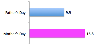
Father's Day versus Mother's Day

Google search terms:


Consumer spending in 2006 (billion)


Average per-person spending in 2010 ($)


Saturday, May 21, 2011

Thy-1 status correlates with fibroblast phenotype

A recent study in collaboration between Selman and Pardo from Mexico, and Hagood of UCSD examined the role of Thy-1, a GPI anchored glycoprotein, in fibroblasts from IPF. They found that Thy-1 negative fibroblasts:
1. Smaller in size;
2. Increased proliferation;
3. Enhanced migration;
4. Enhanced contration;
5. Expression of MMP9 in response to TGFbeta1.

The study is consistent with Hagood's previous reports suggesting Thy-1 negative fibroblasts represent a subset of fibroblasts with elevated activation in fibrotic lung.

Other studies suggested that fibrotic fibroblasts appear to grow slower than normal fibroblasts [2], or there is no difference in growth rate between fibrotic and normal fibroblasts [3].


References
[1] Ramírez G, Hagood JS, Sanders Y, Ramírez R, Becerril C, Segura L, Barrera L, Selman M, Pardo A. Absence of Thy-1 results in TGF-β induced MMP-9 expression and confers a profibrotic phenotype to human lung fibroblasts. Lab Invest. 2011 May 16. [Epub ahead of print] PubMed PMID: 21577212.
[2] Ramos C, Montaño M, García-Alvarez J, Ruiz V, Uhal BD, Selman M, Pardo A. Fibroblasts from idiopathic pulmonary fibrosis and normal lungs differ in growth  rate, apoptosis, and tissue inhibitor of metalloproteinases expression. Am J Respir Cell Mol Biol. 2001 May;24(5):591-8. PubMed PMID: 11350829.
[3] 1: Mio T, Nagai S, Kitaichi M, Kawatani A, Izumi T. Proliferative characteristics of fibroblast lines derived from open lung biopsy specimens of patients with IPF  (UIP). Chest. 1992 Sep;102(3):832-7. PubMed PMID: 1516411.

Thursday, May 19, 2011

No EMT in lung fibrosis

In ATS 2011, a study from Dr. Brigid Hogan's laboratory suggests that mature AT2 cells appear to be capable of clonal proliferation and differentiation into Type 1 alveolar epithelial cells. However, there is no evidence that the lineage-labeled AT2 cells differentiate into mesenchymal cells using an inducible Sftpc-CreER "knock-in" mouse line in combination with a R26R-TomatoRed reporter allele. This study is against a popular hypothesis that the fibroblasts involved in the pathogenesis of IPF derive, in part, from AT2 cells, through EMT [1].

The study is consistent with other studies that did not find evidence for a contribution of EMT to kidney fibrosis [2, 3] or liver fibrosis [4, 5].


References:
[1] Barkauskas, C, et al., Lineage Tracing Of Mature Type 2 Alveolar Epithelial Cells Reveals New Insights Into Alveolar Maintenance And Repair. Am J Respir Crit Care Med 183;2011:A6347
[2] Li L, et al. Autophagy is a component of epithelial cell fate in obstructive uropathy. Am J Pathol. 2010;176(4):1767–1778.
[3] Humphreys BD, et al. Fate tracing reveals the pericyte and not epithelial origin of myofibroblasts in kidney fibrosis. Am J Pathol. 2010 Jan;176(1):85-97. Epub 2009 Dec 11. PubMed PMID: 20008127; PubMed Central PMCID:PMC2797872.
[4] Scholten D, et al. Genetic labeling does not detect epithelial-to-mesenchymal transition of cholangiocytes in liver fibrosis in mice. Gastroenterology. 2010 Sep;139(3):987-98. Epub 2010 Jun 20. PubMed PMID: 20546735; PubMed Central PMCID: PMC2930026
[5] Taura K, et al. Hepatocytes do not undergo epithelial-mesenchymal transition in liver fibrosis in mice. Hepatology. 2010 Mar;51(3):1027-36. PubMed PMID: 20052656; PubMed Central PMCID: PMC2906231.

Pirfenidone has a benifit for IPF

A new study published in Lancet online reported the results from two randomized trials that pirfenidone has a favourable benefit risk profile and represents an appropriate treatment option for patients with idiopathic pulmonary fibrosis [1]. These two trials were designed to test pirfenidone 2403 mg/day and 1197 mg/day. In one trials, high-dose pirfenidone significantly reduced the decline in % pred FVC. A consistent pirfenidone treatment effect was found up to week 48. A significant reduction in the decline from baseline to week 72 in 6MWT distance was observed in patients with pirfenidone treatment. Pirfenidone prolonged progression-free survival by 26% compared with placebo.

These trials are encouraging in gaining FDA approval use of pirfenidone in the US.


Fig 1A.


 [1] Noble PW, et al. Pirfenidone in patients with idiopathic pulmonary fibrosis
(CAPACITY): two randomised trials. Lancet. 2011 May 13. [Epub ahead of print]
PubMed PMID: 21571362.


MUC5B is a susceptible gene for IPF

A recent study by Dr. David Schwartz of University of Colorado showed that a SNP in the promoter region of MUC5B is strongly associated with FPF and IPF [1]. The odds ratios for disease among subjects who were heterozygous and those who were homozygous for the minor allele of this SNP (rs35705950) were 6.8 and 20.8, respectively, for FPF. The odds ratios for IPF were similar (9.0 and 21.8). MUC5B expression in the lung was 14.1 times as high in subjects with IPF as in those healthy donors.

The significance of the study, along with previous reports that variants in SFTPA, SFTPB, SFTPC, ABCA1, and ABCA3 were found in patients with pulmonary fibrosis, points to a plausible notion that the dysregulated secretion of lung surfactants and mucins by airway and alveolar epithelial cells leads to endoplasmic-reticulum (ER) stress. ER stress is found in alveolar epithelial cells in IPF [2]. However, we do not know the dysregulation of these proteins is duo to a common set of transcription factors or to epigenetic regulations.

[1] Seibold MA,  et al. A common MUC5B promoter polymorphism and pulmonary fibrosis. N Engl J Med. 2011 Apr 21;364(16):1503-12. PubMed PMID: 21506741.

[2] Korfei M, et al. Epithelial endoplasmic reticulum stress and apoptosis in sporadic idiopathic pulmonary fibrosis. Am J Respir Crit Care Med. 2008 Oct 15;178(8):838-46. Epub 2008 Jul 17. PubMed PMID: 18635891; PubMed Central PMCID: PMC2566794.

Thursday, April 14, 2011

Methylation and Kidney Fibrosis

A team of scientists led by Drs. Kalluri and Zeisberg of Harvard suggested that demethylation reduced kidney fibrosis in mice.

The potential mechanisms of this effect may be various.  They found that a RAS inhibitor, RASAL1, is hypermethylated in renal fibrosis.


Nat Med. 2010 May;16(5):544-50. Epub 2010 Apr 25.
Methylation determines fibroblast activation and fibrogenesis in the kidney.
Bechtel W, McGoohan S, Zeisberg EM, Müller GA, Kalbacher H, Salant DJ, Müller CA, Kalluri R, Zeisberg M.
Division of Matrix Biology, Department of Medicine, Beth Israel Deaconess Medical Center and Harvard Medical School, Boston, Massachusetts, USA.
Comment in:
Nat Med. 2010 May;16(5):523-5.
Kidney Int. 2010 Sep;78(5):430.
Abstract
Fibrogenesis is a pathological wound repair process that fails to cease, even when the initial insult has been removed. Fibroblasts are principal mediators of fibrosis, and fibroblasts from fibrotic tissues fail to return to their quiescent stage, including when cultured in vitro. In a search for underlying molecular mechanisms, we hypothesized that this perpetuation of fibrogenesis is caused by epigenetic modifications. We demonstrate here that hypermethylation of RASAL1, encoding an inhibitor of the Ras oncoprotein, is associated with the perpetuation of fibroblast activation and fibrogenesis in the kidney. RASAL1 hypermethylation is mediated by the methyltransferase Dnmt1 in renal fibrogenesis, and kidney fibrosis is ameliorated in Dnmt1(+/-) heterozygous mice. These studies demonstrate that epigenetic modifications may provide a molecular basis for perpetuated fibroblast activation and fibrogenesis in the kidney.
PMID: 20418885 [PubMed - indexed for MEDLINE]

Wednesday, April 13, 2011

Fstl1 is a BMP4 signaling antagonist in controlling mouse lung development

Proc Natl Acad Sci U S A. 2011 Apr 11. [Epub ahead of print]

Follistatin-like 1 (Fstl1) is a bone morphogenetic protein (BMP) 4 signaling antagonist in controlling mouse lung development.

Model Animal Research Center, Nanjing University, Nanjing 210061, China.

Abstract

Lung morphogenesis is a well orchestrated, tightly regulated process through several molecular pathways, including TGF-β/bone morphogenetic protein (BMP) signaling. Alteration of these signaling pathways leads to lung malformation. We investigated the role of Follistatin-like 1 (Fstl1), a secreted follistatin-module-containing glycoprotein, in lung development. Deletion of Fstl1 in mice led to postnatal lethality as a result of respiratory failure. Analysis of the mutant phenotype showed that Fstl1 is essential for tracheal cartilage formation and alveolar maturation. Deletion of the Fstl1 gene resulted in malformed tracheal rings manifested as discontinued rings and reduced ring number. Fstl1-deficient mice displayed septal hypercellularity and end-expiratory atelectasis, which were associated with impaired differentiation of distal alveolar epithelial cells and insufficient production of mature surfactant proteins. Mechanistically, Fstl1 interacted directly with BMP4, negatively regulated BMP4/Smad1/5/8 signaling, and inhibited BMP4-induced surfactant gene expression. Reducing BMP signaling activity by Noggin rescued pulmonary atelectasis of Fstl1-deficient mice. Therefore, we provide in vivo and in vitro evidence to demonstrate that Fstl1 modulates lung development and alveolar maturation, in part, through BMP4 signaling.
PMID: 21482757 [PubMed - as supplied by publisher]


PMID: 21482757 [PubMed - as supplied by publisher]


Tuesday, April 12, 2011

G2/M unrest and kidney fibrosis

Epithelial cell cycle arrest in G2/M mediates kidney fibrosis after injury.
Yang L, Besschetnova TY, Brooks CR, Shah JV, Bonventre JV.

Department of Medicine, Renal Division, Brigham and Women's Hospital, Harvard Medical School, Boston, Massachusetts, USA.
Comment in:

Nat Med. 2010 May;16(5):523-5.
Abstract
Fibrosis is responsible for chronic progressive kidney failure, which is present in a large number of adults in the developed world. It is increasingly appreciated that acute kidney injury (AKI), resulting in aberrant incomplete repair, is a major

contributor to chronic fibrotic kidney disease. The mechanism that triggers the fibrogenic response after injury is not well understood. In ischemic, toxic and obstructive models of AKI, we demonstrate a causal association between epithelial cell cycle G2/M arrest and a fibrotic outcome. G2/M-arrested proximal tubular cells activate c-jun NH(2)-terminal kinase (JNK) signaling, which acts to upregulate profibrotic cytokine production. Treatment with a JNK inhibitor, or bypassing the G2/M arrest by administration of a p53 inhibitor or the removal of the contralateral kidney, rescues fibrosis in the unilateral ischemic injured kidney. Hence, epithelial cell cycle arrest at G2/M and its subsequent downstream signaling are hitherto unrecognized therapeutic targets for the prevention of fibrosis and interruption of the accelerated progression of kidney disease.

http://www.ncbi.nlm.nih.gov/pubmed/20436483
[PubMed - indexed for MEDLINE]


pirfenidone approved for idiopathic pulmonary fibrosis in EU

Nat Biotechnol. 2011 Apr;29(4):301.

p38 kinase inhibitor approved for idiopathic pulmonary fibrosis.

Moran N.

PMID: 21478838 [PubMed - in process]


{beta}-Arrestin Deficiency Protects Against Pulmonary Fibrosis in Mice and Prevents Fibroblast Invasion of Extracellular Matrix.

Sci Transl Med. 2011 Mar 16;3(74):74ra23.
{beta}-Arrestin Deficiency Protects Against Pulmonary Fibrosis in Mice and Prevents Fibroblast Invasion of Extracellular Matrix.
Lovgren AK, Kovacs JJ, Xie T, Potts EN, Li Y, Foster WM, Liang J, Meltzer EB, Jiang D, Lefkowitz RJ, Noble PW.

Division of Pulmonary, Allergy, and Critical Care Medicine, Department of Medicine, Duke University Medical Center, Durham, NC 27710, USA.
Abstract
Idiopathic pulmonary fibrosis is a progressive disease that causes unremitting extracellular matrix deposition with resulting distortion of pulmonary architecture and impaired gas exchange. β-Arrestins regulate G protein (heterotrimeric guanine nucleotide-binding protein)-coupled receptors through receptor desensitization while also acting as signaling scaffolds to facilitate numerous effector pathways. Here, we examine the role of β-arrestin1 and β-arrestin2 in the pathobiology of pulmonary fibrosis. In the bleomycin-induced mouse lung fibrosis model, loss of either β-arrestin1 or β-arrestin2 resulted in protection from mortality, inhibition of matrix deposition, and protected lung function. Fibrosis was prevented despite preserved recruitment of inflammatory cells and fibroblast chemotaxis. However, isolated lung fibroblasts from bleomycin-treated β-arrestin-null mice failed to invade extracellular matrix and displayed altered expression of genes involved in matrix production and degradation. Furthermore, knockdown of β-arrestin2 in fibroblasts from patients with idiopathic pulmonary fibrosis attenuated the invasive phenotype. These data implicate β-arrestins as mediators of fibroblast invasion and the development of pulmonary fibrosis, and as a potential target for therapeutic intervention in patients with idiopathic pulmonary fibrosis.

PMID: 21411739 [PubMed - in process]